Rutgers logo

Enter a Search Term

Rutgers logo
  • Rutgers.edu
Rutgers logo
  • Home
  • Office
  • Areas of Excellence
    • Brain Injury & Stroke
    • Center for Cell Signaling
    • Cardiovascular Biology
    • Immunology & Infectious Diseases
    • Neurosciences
    • Stem Cells & Regeneration
  • Core Facilities
    • Center for Advance Proteomics Research
    • Cellular Imaging & Histology
    • Flow Cytometry & Immunology Core Laboratory
    • Genomics at NJMS
    • Molecular & Genomics Informatics Core
    • Clinical Research Unit (CRU)
    • Additional Research Core Facilities
      • Biospherix™ Hypoxia Chamber
      • Comparative Medicine Resources
      • Irradiation Facility
      • Shared Resources at Rutgers Cancer Institute of New Jersey
      • Shared Facilities at the Public Health Research Institute
  • Research Centers
    • Center for Immunity & Inflammation
    • Center for Cell Signaling
    • Public Health Research Institute
    • Regional Biocontainment Laboratory (RBL)
    • Center for Emerging Pathogens
  • OCRA
    • Forms
    • UH Medical Records Access
    • UH Report Request
    • Contact Us
    • Additional Resources
      • NJMS CRU
      • NJ ACTS
      • RBHS Clinical Trials Office
      • Human Subjects Protection Program
      • Research Administration & Proposal Submission System (RAPSS)
  • Grants & Contracts
    • Research Administration
      • Research Administrative Support Team
      • Grant Submissions
      • Sponsors
      • Funding Opportunities
    • Intramural Grants Program
      • NMS Core Facilities Matching Funds Program
      • RBHS Bridge Grants Program
  • Training Programs
    • Summer Student Research Program
    • High School Internship Program
    • Unpaid Internship Program
    • MD with Thesis Program
  • Links
    • University Procurement Services
    • Office for Research Innovation Ventures
    • University Policies
    • University Finance & Administration
    • Research & Sponsored Programs
    • IT@NJMS
    • School of Graduate Studies
    • University Libraries
Rutgers logo
  • Home
  • Office
  • Areas of Excellence
  • Core Facilities
  • Research Centers
  • OCRA
  • Grants & Contracts
  • Training Programs
  • Links
  • Rutgers.edu

Blood Chromosome Analysis

Blood Chromosome Analysis
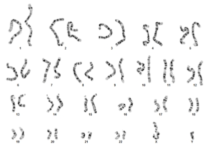
Image of chromosomes

Background:

Cells from peripheral blood are used most often in chromosome analysis, although other cell types can also yield good results. Cells from peripheral blood have several advantages: they are easy to collect, can be cultured rapidly (in three days or less), and only a few drops of blood are necessary for analysis.


Indications for Testing:

Clinical uses of stimulated peripheral blood cultures include:

  • Determination of the constitutional karyotype of patients requiring genetic diagnosis or genetic counseling;
  • Determination of the constitutional karyotype of families of individuals with chromosome abnormalities to determine carrier status;
  • Determination of the constitutional karyotype of leukemia patients who have chromosome abnormalities in their bone marrow;
  • Assessment of chromosome damage in individuals exposed to environmental hazards.


Sample Requirements:

Five milliliters of whole blood collected by venipuncture into a green top sodium heparin tube. In newborns, 1-2 mls of blood may be sufficient.


Specimen Handling:

Blood may be stored at room temperature overnight, refrigerated up to three days, but never frozen. Blood may also be mailed by overnight courier at ambient temperature. Blood drawn more than five days prior to receipt, or in the wrong tube (such as lithium heparin), is unacceptable.


Procedure:

The blood is cultured for 72 hours in culture media with fetal bovine serum, L-glutamine and PHA (phytohemagglutanin) at 5% CO2 and 37C. After culture, blood is exposed to colcemid (to collect mitotic cells), hypotonic solution and fixative, and the cell suspension is dropped on a slide. Slides are G-banded, and 20 cells and two karyotypes are prepared from each routine case.


Interpretation:

Peripheral blood analysis will detect numerical chromosomal abnormalities, such as trisomies, monosomies and chromosomal mosaicism; structural abnormalities such as translocations, inversions, derivative chromosomes and additional abnormal chromosomes. Deletions and rearrangements on the molecular level cannot be seen. The origin and structure of derivative and structurally abnormal chromosomes can be deduced by following G-banding analysis by fluorescent in situ hybridization (FISH).

Microdeletion syndromes can be detected by FISH analysis (see FISH section).


Results:

Results are presented according to the international standards for chromosomal nomenclature (ISCN), and a full explanation of the karyotype and clinical implications is provided. Any abnormal or variant results are followed by genetic counseling.

Site Footer

Rutgers logo

QUICK LINKS

  • George F Smith Library
  • Centers & Institutes
  • Make a Gift
  • Nondiscrimination Statement
  • RBHS
  • Rutgers Health
  • Rutgers University

ABOUT US

  • Administrative Office
  • Leadership Team
  • Contact GCA Staff
  • Contact Compliance Staff
  • Contact OCRA Staff

AFFILIATIONS

  • University Hospital
  • RWJBarnabas Health
  • Veterans Health Administration
  • Bergen New Bridge Medical Center
  • Trinitas Regional Medical Center

CONTACT US

Medical Science Building
185 South Orange Avenue
Newark, NJ 07103
(732) 445-info (4636)
njmsmarketing@njms.rutgers.edu
Google Map

Follow Us

Rutgers is an equal access/equal opportunity institution. Individuals with disabilities are encouraged to direct suggestions, comments, or complaints concerning any accessibility issues with Rutgers websites to accessibility@rutgers.edu or complete the Report Accessibility Barrier / Provide Feedback form.

Copyright ©2025, Rutgers, The State University of New Jersey. All rights reserved. Contact webmaster