Cellular Imaging and Histology Core
Cellular Imaging & Histology
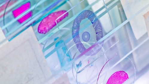
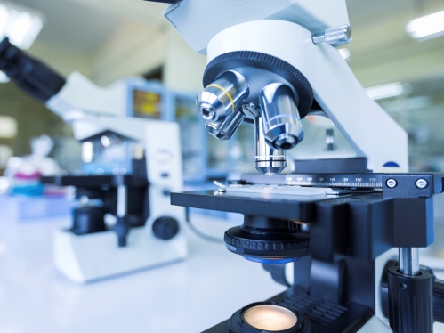
The Cellular Imaging and Histology Core integrates the preparation of research tissue specimens for microscopic investigation with imaging capabilities for brightfield and fluorescent microscopy. The core staff is able to prepare research tissue specimens for Histological sectioning utilizing both frozen and paraffin embedding techniques. The facility maintains microtomes and cryostats for the preparation of tissue sections. The Core staff can provide fee-for-service preparation of mounted sections on glass slides or these instruments can be rented for operation by users of the core. The Core maintains several instruments for the basic documentary capture of microscope slides and houses several high end fluorescent microscopy systems, including the Leica Stellaris 8 tau-STED Super Resolution Microscope, the Nikon A1R+ HD Confocal Microscope, and the Nikon Ti2 HCA Inverted Fluorescence Microscope.
Personnel

Luke Fritzky

Dongfang Liu, Ph.D., MBBS

Joel Pierre
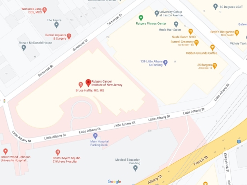
Location
Rutgers, The State University of New Jersey
New Jersey Medical School
Cancer Institute of New Jersey at University Hospital
205 South Orange Ave. Room G-1105
Newark, NJ 07103
Hours of Operation
Monday through Friday 9:00 AM - 5:00 PM