Advanced Preclinical Imaging Laboratory Equipment List
Advanced Preclinical Imaging Laboratory Equipment List
Accordion Content
-
Session Internal Price External Price Self Use TBD TBD Assisted Use/Training TBD TBD 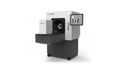
(https://www.milabs.com/preclinical-imaging-systems/)
The Milab Vector6 is a versatile imaging system that integrates Positron Emission Tomography (PET), Single Photon Emission Computed Tomography (SPECT), and Computed Tomography (CT) capabilities. This allows for high-resolution anatomical imaging (CT), functional imaging (PET), and molecular imaging (SPECT).
-
Session Internal Price External Price Self Use TBD TBD Assisted Use/Training TBD TBD 
https://www.revvity.com/product/ivis-spectrum-in-vivo-imaging-system
The IVIS Spectrum provides highly sensitive bioluminescence and fluorescence imaging for small animals. It enables real-time, non-invasive monitoring of disease progression, tumor growth, gene expression, and therapeutic response. With its multispectral capabilities, it enables the spectral unmixing of multiple fluorophores. -
Session Internal Price External Price Self Use TBD TBD Assisted Use/Training TBD TBD 
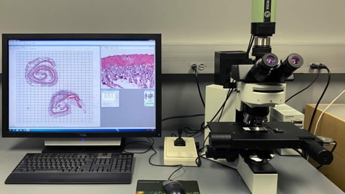
Our 1T MRI scanner offers high-quality preclinical imaging with a focus on anatomical and functional characterization. This system is ideal for a wide range of applications, including structural imaging, and diffusion-weighted imaging (DWI), among others.
-
Session Internal Price External Price Self Use TBD TBD Assisted Use/Training TBD TBD 
The skyscan 1172 is a cutting-edge computed tomography system designed for ex vivo imaging applications, with high spatial resolution, it enables detailed anatomical imaging for small animals, biomaterials among others.
-
Session Internal Price External Price Self Use TBD TBD Assisted Use/Training TBD TBD 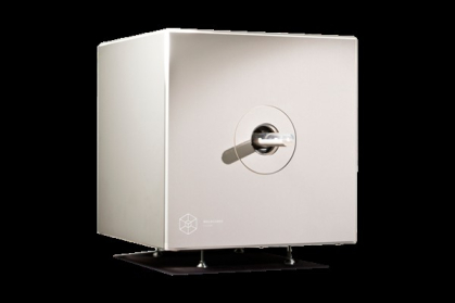
The Molecubes X-CUBE is a compact, high-resolution, low-dose micro-CT imaging system optimized for small animal imaging. It provides rapid 3D anatomical visualization and is located within the BLS3 facility at RBL–Rutgers Newark, supporting translational and infectious disease research under controlled biosafety conditions.
-
Research Focus and Disease Models
Our lab utilizes advanced imaging technologies and radiochemistry to investigate diverse disease models in preclinical research. Below is an overview of our key areas of focus and how we apply each imaging modality:
Oncology
- Tumor Characterization:
- PET/SPECT: Radiotracers reveal tumor metabolism and molecular markers.
- CT: Provides precise 3D anatomical images to locate tumors and monitor their size.
- IVIS Spectrum: Enables non-invasive optical imaging of tumor growth, metastasis, and therapeutic response using bioluminescent and fluorescent probes.
- Treatment Response: Tracking metabolic, optical, and anatomical changes with PET, CT, and IVIS Spectrum.
- Radiotherapeutics: Development and evaluation of targeted radioactive treatments.
Disease Models:
- Xenograft and genetically engineered mouse models.
Neuroimaging
- Brain Structure and Connectivity:
- MRI: Detailed imaging of brain anatomy and neural pathways via diffusion-weighted imaging (DWI).
- CT: Structural imaging for calcifications or hemorrhage detection when needed.
- Neuroreceptor Imaging: PET/SPECT imaging of neurotransmitter systems.
- Neurodegeneration: Imaging pathological protein accumulation and inflammation.
- IVIS Spectrum: Facilitates studies on neuronal gene expression and neuroinflammation using reporter systems.
Disease Models:
- Transgenic models of Alzheimer’s, Parkinson’s, stroke, and brain injury.
Cardiovascular Imaging
- Cardiac Function and Structure:
- CT: Offers high-resolution visualization of coronary arteries, vascular calcifications, and cardiac anatomy, useful for assessing structural abnormalities.
- Molecular and Functional Imaging:
- PET: Measures myocardial perfusion, metabolism, and viability using specialized radiotracers to assess heart muscle health and detect ischemia.
- SPECT: Evaluates blood flow and cardiac function, commonly used for myocardial perfusion imaging to diagnose coronary artery disease.
- IVIS Spectrum: Optical imaging for angiogenesis and targeted fluorescent probes.
- Ischemia and Infarction: Integrated imaging approach to monitor heart tissue damage, repair processes, and therapeutic interventions.
Disease Models:
- Preclinical models of myocardial infarction, heart failure, ischemic heart disease, and atherosclerosis.
Bone and Dental Imaging
- CT: High-resolution 3D imaging of bone microarchitecture and dental structures to study bone health, fractures, implant integration, and dental disease progression.
- Applications: Assessment of osteoporosis models, bone healing, dental caries, and periodontal disease in small animals.
Disease Models:
- Osteoporosis, fracture healing, and dental disease models in rodents.
Obesity and Metabolic Disease
- CT: Non-invasive imaging to quantify fat distribution (visceral and subcutaneous fat), liver fat infiltration, and organ changes related to obesity and metabolic syndrome.
- Complementary Modalities: PET imaging to study metabolic activity in adipose tissue and related organs.
Disease Models:
- Diet-induced obesity and genetically modified obesity models in rodents.
Lung Imaging
- CT: Detailed anatomical imaging of lung structure for detecting inflammation, fibrosis, tumors, or emphysema. High-resolution CT allows quantitative assessment of lung volumes and pathology.
- Molecular Imaging: PET/SPECT tracers to visualize inflammation and infection processes.
- IVIS Spectrum: Optical tracking of inflammatory cell infiltration and infection progression.
Disease Models:
- Models of pulmonary fibrosis, chronic obstructive pulmonary disease (COPD), lung cancer, and infection.
Molecular Imaging
- Radiotracer Development: Creation of novel PET and SPECT tracers targeting disease-specific molecular pathways.
- Targeted Imaging: Combining molecular imaging with CT for precise localization and quantification of biological processes like hypoxia, apoptosis, and immune responses.
- IVIS Spectrum: Advanced optical imaging for hypoxia, apoptosis, immune responses, and real-time gene expression studies.
- Tumor Characterization: